 The cornea, or clear covering at the front of your eye, has two important functions. It helps protect the other structures of your eye from foreign debris and helps focus light entering your eye.
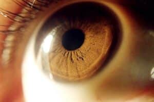
The shape of your cornea determines how light is focused on the retina in the back of your eye. A normal, healthy cornea has a round shape like a dome, and focuses light directly on the retina. The retina receives the light, converts it into signals and transmits the signals to the brain for visual recognition.
Corneas that develop a degenerative disease called keratoconus thin and bulge abnormally, taking on a more cone-like shape. This irregularity is known as ectasia, and it interrupts the focusing of light rays, causing blurry, distorted vision that cannot be corrected with glasses or contact lenses.
The team at Vista Eye Specialists has special expertise in the management of corneal disorders, including keratoconus. We have seen particularly excellent results with scleral contact lenses and rigid gas permeable contact lenses. Corneal crosslinking, another treatment option, is available at our partner practices in northern Virginia and Richmond.
Keratoconus: Overview
Corneal Crosslinking for Keratoconus
 Corneal crosslinking is designed to strengthen the collagen crosslinks in the inner layers of the cornea. Collagen fiber crosslinks function as structural support for the cornea, but corneas affected by keratoconus do not have adequate crosslinks.
Corneal crosslinking involves applying a combination of ultraviolet light and riboflavin drops to the eyes, causing new collagen crosslinks to develop. This stabilizes the cornea and prevents it from bulging further. The treatment itself can be performed at our office and takes approximately one hour.
2 Approaches to Corneal Crosslinking
There are two approaches to corneal crosslinking. In epithelium-off crosslinking, the outer layer of the cornea (called the epithelium) is removed so the riboflavin drops can soak into the deeper layers of the corneal tissue. In epithelium-on crosslinking, the outer layer of cells is left intact.
Corneal Crosslinking Recovery
After treatment, side effects like sensitivity to light or the sensation of a foreign body in the eye can occur. In epithelium-off crosslinking, there may be slightly more discomfort as the cornea heals. But these aftereffects are mild and temporary. Medicated eyedrops must be used after treatment to prevent infection and promote healing.
Corneal crosslinking works best in cases of mild to moderate keratoconus. It cannot cure the disease, but it can stop it from progressing. Some patients respond to a single corneal crosslinking treatment session, and others need several treatment sessions.
Post-LASIK Ectasia
Keratoconus is not the only factor that can cause corneal ectasia. Rarely, corneal ectasia can be a complication of laser eye surgery procedures like LASIK or PRK. In patients with naturally thin corneas, high myopia or irregular astigmatism, removing corneal tissue with a laser during LASIK or PRK weakens the cornea permanently. Luckily, corneal crosslinking can strengthen the corneas of patients affected by post-LASIK or post-PRK ectasia.
What are the symptoms of keratoconus?
Keratoconus weakens the cornea, the clear layer of tissue at the front of the eye. The cornea thins and bulges out, creating a cone shape as it weakens.
Because your corneas isare essential for focusing light onto the retina, this change can trigger some frustratingfrustrating vision problems, including:
- Gradual decline in visual clarity
- Blurry or distorted vision
- Light sensitivity
- Increased glare
- Frequent prescription changes (glasses or contact lenses)
- Double vision
As keratoconus progresses, these symptoms can worsen and reduce your quality of life. In advanced cases, vision can deteriorate significantly, making it difficult to see well, even with glasses or contact lenses.
What causes keratoconus?
 The exact cause of keratoconus isn’t known, but experts believe that genetics and environment may both play a role. Some people may inherit it from their parents, but not everyone with a family history of the condition develops it themselves.
Who is at risk for keratoconus?
Keratoconus often begins in one’s teenage years or early adulthood. Some known risk factors include:
- Family history of keratoconus
- Chronic eye rubbing
- Eye allergies
- Connective tissue disorders, such as Ehlers-Danos syndrome
Understanding your risk factors can be crucial for effectively detecting and managing symptoms.
What are the complications of keratoconus?
As keratoconus progresses, complications can include:
- Scarring: Thinning of the cornea can cause scarring, especially in advanced cases.
- Hydrops: This is a sudden swelling in the cornea due to fluid leakage, which can cause severe vision impairment.
- Difficulty with Contact Lenses: In advanced keratoconus, contact lenses may become uncomfortable and require specialized lenses.
Although it rarely causes complete blindness, vision loss may progress to the point of legal blindness in some people with keratoconus.
What is corneal cross-linking, and how does it treat keratoconus?
 Corneal cross-linking is a minimally invasive office-based procedure that improves cornea strength using a combination of UV light and a riboflavin (vitamin B2) solution.
This process helps to stabilize the corneal shape, slowing or halting the progression of keratoconus. Although it may not reverse the damage, cross-linking can prevent further vision loss and help many patients avoid corneal transplants.
When is a corneal transplant necessary for keratoconus?
For patients with advanced keratoconus who do not respond to other treatments, our eye surgeons may recommend a corneal transplant.
Surgery involves replacing the damaged cornea with healthy donor tissue to restore vision.
Corneal transplants can have a high success rate. Recovery can be extensive, requiring months to see optimal results; however, for patients whose vision is at risk, the surgery can be life changing.
How much does keratoconus treatment cost?
Costs vary depending on the type of treatment:
- Specialty Contact Lenses: Often needed for mild to moderate keratoconus, these special lenses can cost more than standard lenses. Examples include rigid gas-permeable contacts (hard lenses), hybrid lenses, and scleral lenses that vault over the cornea. Some insurance plans may cover the cost if they’re considered medically necessary.
- Corneal Cross-Linking: As an FDA-approved treatment, cross-linking is covered by many insurance providers, though coverage details vary by plan.
- Corneal Transplants: Insurance plans often cover corneal transplants for patients with severe keratoconus. However, plan details and associated costs vary.
We recommend contacting your insurance provider to understand the coverage for keratoconus treatments.